Utility of [18F]FDG-PET/CT for Detecting activity in Schnitzler Syndrome
- 1. Department of Nuclear Medicine, Ramón y Cajal Universitary Hospital, Spain
CITATION
Martinez-Lorca A, Ajuria-Illarramendi O, Diez MD (2025) Utility of [18F]FDG-PET/CT for Detecting activity in Schnitzler Syndrome. J Radiol Radiat Ther 13(1): 1108.
INTRODUCTION
Schnitzler síndrome [1], is a rare autoinflammatory disorder characterized by a combination of symptoms that include recurrent urticaria (hives), fever, joint pain, and the presence of a monoclonal IgM paraprotein in the blood. Diagnosis is primarily based on clinical criteria and laboratory findings, including the detection of the paraprotein and the exclusion of other diseases. Treatment often involves the use of IL-1 inhibitors.
CASE REPORT
A 49-year-old woman has been under follow-up since 2021 for recurrent urticaria, prolonged low-grade fever, asthenia, and osteoarticular pain. She presents with monoclonal IgM paraprotein and associated positive cryoglobulinemia, without signs of vasculitis, hypocomplementemia, or renal involvement, and no presence of Raynaud’s phenomenon or ischemic events. Bone marrow aspiration was negative for lymphoproliferative disorder.
At the time of diagnosis, a CT skull and a thoraco- abdominopelvic CT were requested to rule out organic causes, which showed mild splenomegaly and subcentimeter retroperitoneal lymphadenopathy (Figure 1).
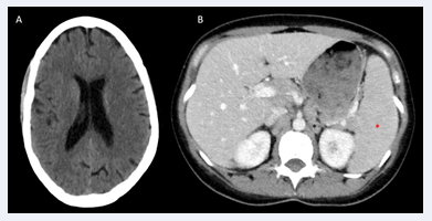
Figure 1: A, axial CT image of the skull without alterations; B, axial thoraco-abdominopelvic CT image showing subcentimeter retroperitoneal lymphadenopathy (black asterisk) and splenomegaly (red asterisk).
The patient was diagnosed with Schnitzler syndrome and started treatment with subcutaneous anakinra [2], showing very favorable clinical evolution from the cutaneous, systemic, and articular perspectives.
In December 2023, the patient presented with a PCR of 10.8 mg/L, elevated serum IgM (818,000 mg/dL, previously 551,000 mg/dL), and persistence of cryoglobulinemia (cryoprecipitate represents 6% of total volume) without clear clinical, cutaneous, or articular worsening.
Given the absence of symptoms and the analytical data, a [18F]FDG-PET-CT study was decided, which revealed hepatosplenomegaly [3], inversion of the hepatic-splenic metabolic gradient, and hypermetabolic lymphadenopathy related to reactivation of her underlying process (Figure 2).
![Figure 2 [18F]FDG-PET/CT whole-body study. A, MIP image; B, axial CT image; C, axial PET image; D, axial PET/CT fusion image. The study shows multiple infradiaphragmatic lymphadenopathies (black asterisk) and splenomegaly with inversion of the metabolic gradient (red asterisk).](https://www.jscimedcentral.com/public/assets/images/uploads/image-1749713608-1.PNG )
Figure 2: [18F]FDG-PET/CT whole-body study. A, MIP image; B, axial CT image; C, axial PET image; D, axial PET/CT fusion image. The study shows multiple infradiaphragmatic lymphadenopathies (black asterisk) and splenomegaly with inversion of the metabolic gradient (red asterisk).
Given the presence of these findings and based on the previous favorable response to IL-1 blockade treatment, the treatment was modified to subcutaneous canakinumab, with the patient showing stability and improvement in IgM levels (287.00 mg/dL).
DISCUSSION
The [18F]FDG-PET/CT [4], is a consolidated functional imaging technique that has demonstrated its role in the field of inflammatory diseases. While it does not seem to be routinely useful for the diagnosis and follow-up of Schnitzler síndrome [5], it should be considered in cases where clinical/analytical worsening is suspected for the assessment of unknown inflammatory activity or possible evolution to a lymphoproliferative process.
This imaging technique serves as a non-invasive and highly sensitive diagnostic tool in Schnitzler Syndrome aiding in disease monitoring and personalized treatment strategies.
One of the main challenges in diagnosing Schnitzler syndrome is differentiating it from other autoinflammatory or hematologic disorders. [18F]FDG-PET/CT aids in this process by highlighting areas of increased metabolic activity, which correspond to inflammation. Several studies have demonstrated the utility of this imaging technique in detecting systemic inflammatory involvement [6], in Schnitzler syndrome patients.
[18F]FDG-PET/CT findings often reveal hypermetabolic activity in the bone marrow, lymph nodes, and spleen. This is particularly useful in distinguishing Schnitzler syndrome from malignancies such as lymphoma. Additionally, [18F] FDG-PET/CT can help exclude infectious or neoplastic conditions that might mimic Schnitzler syndrome.
[18F]FDG-PET/CT could be an important technique in monitoring disease [7], progression and evaluating response to therapy. The primary treatment for Schnitzler syndrome is IL-1 inhibition (e.g., anakinra or canakinumab). [18F]FDG-PET/CT imaging can objectively assess changes in metabolic activity following treatment initiation, thereby guiding clinical decision-making.
Patients with Schnitzler syndrome who respond well to IL-1 inhibitors typically show a marked reduction in FDG uptake in previously affected regions. Conversely, persistent metabolic activity despite treatment may indicate inadequate therapeutic response or the need for alternative interventions.
[18F]FDG-PET/CT plays a crucial role in the diagnostic workup, disease monitoring, and therapeutic assessment of Schnitzler syndrome. By providing a non-invasive and sensitive method to visualize systemic inflammation, improves diagnostic accuracy, differentiates from malignancies, and assists in evaluating treatment efficacy. As research continues, this imaging modality may become even more integral in managing patients with Schnitzler syndrome and other autoinflammatory diseases.
REFERENCES
- Gallo J, Paira S. Síndrome de Schnitzler. Reumatol Clin. 2015; 11: 124-125.
- Hidalgo-García Y, García-Fernández E, Palacio-Aller L, Gonzalvo P. Schnitzler Syndrome with response to Anakinra Monotherapy: 7 Years of Follow-up. Actas Dermosifiliogr. 2017; 108: 956-958.
- Lipsker D. The Schnitzler síndrome. Orphanet J Rare Dis. 2010; 5: 38.
- Riemer HJA, Slart, Andor WJM, Glaudemans, Panithaya Chareonthaitawee, Giorgio Treglia, Florent L Besson, Thorsten A Bley, et al. FDG-PET/CT(A) imaging in large vessel vasculitis and polymyalgia rheumatica: joint procedural recommendation of the EANM, SNMMI, and the PET Interest Group (PIG), and endorsed by the ASNC. Eur J Nucl Med Mol Imaging. 2018; 45: 1250-1269.
- Alix L, Néel A, Cador B, Smail A, Serratrice J, Closs-Prophette F, et al. Diagnostic value of 18-F fluorodeoxyglucose PET/CT and bone scan in Schnitzler syndrome. Autoimmunity. 2019; 52: 264-271.
- Lai N, Tan K, Khan S, Barwick T, Marzola MC, Rubello D, et al. Role of FDG-PET/CT in the diagnosis and management of autoinflammatory syndromes: A review. Clinical Rheumatology. 2020; 39: 1679-1691.
- Glatard A. FDG-PET/CT findings in Schnitzler syndrome: A potential diagnostic and follow-up tool. Eur J Nucl Med Mol Imaging. 2019; 46: 1121-1128.


